Brains are highly adaptable. Neuronal connections are constantly changing, molded by what people experience in their daily lives. New memories, information learned, and new skills spark this dynamic process, causing lasting changes to neuronal circuits. As the saying goes, experience is the best teacher, and it couldn’t be truer for brains.
Along with learning and memory, sensory experiences such as listening to music or appreciating a stunning view also have a similar effect on the brain. Incoming sensory information activates neurons in the cortex, causing long-term modifications to the circuitry depending on what is experienced. This process, called “experience dependent plasticity,” is part of the reason why brains develop differently due to unique individual life experiences.
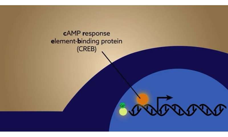
But how does the brain convert relatively short-lasting neuronal activity into the long-term changes driven by sensory experiences? The key lies in specialized proteins called activity-dependent transcription factors. Responding to neuronal activity, these factors activate genes within the cell to translate rapid incoming signaling into slower, lasting changes. Despite the evident importance of activity-dependent transcription to development and long-term plasticity, it was previously impossible to directly monitor transcription factor activity. This was mainly due to the lack of available tools to study the interaction between neuronal activity and transcription factor activation that occurs in an intact, living brain.
In a recently published study in Neuron, scientists in the Yasuda Lab at the Max Planck Florida Institute for Neuroscience (MPFI) have designed and developed novel biosensors that allow the simultaneous study of both sensory-evoked neuronal activity and transcription factor dynamics. Coupling the specialized techniques of two-photon calcium imaging with two-photon fluorescence lifetime imaging (2pFLIM), scientists now have the ability to investigate how transcription factors function in a living brain with single-cell resolution.
https://youtube.com/watch?v=7ofhU7qjX14%3Fcolor%3Dwhite
“Transcription factor activity in the brain isn’t static, it’s a very dynamic process that can occur on the order of hours to days after a sensory experience,” explains Dr. Tal Laviv, research fellow in the Yasuda Lab and leading author of the paper. “Traditional methods of studying these proteins involve freezing brain tissue at a single moment in time. So while these approaches can tell you if a certain factor is activated or not, they aren’t good at capturing how experience shapes transcription factor activity over time. We wanted to develop a new way to study how this process is actually occurring in a living brain, and we chose to study CREB due to its strong involvement in plasticity, learning and memory.”
MPFI scientists started by creating sensitive and specific 2pFLIM biosensors designed to report the direct activity of cAMP response-element binding protein (CREB). Packaging their newly generated sensors using an adeno-associated viral strategy (AAVs), the team then expressed them in a population of neurons within the somatosensory cortex of mice. Changes in environment are known to activate this brain region, and in response, the neurons within modulate numerous signaling molecules including CREB. But little is known about the temporal dynamics of CREB activation after a change to the environment. Using the 2pFLIM CREB sensor, the team chronically monitored CREB activity in the same population of neurons while mice experienced an enriched environment. The enriched environment caused a significant increase in overall CREB activity. Interestingly, when mice were removed from the enriched environment for an extended period of time, CREB activity returned to normal levels, indicating sensory experience as a driver for the sustained activity.
Next, MPFI scientists sought to unravel how sensory experiences and neuronal activation shape CREB activity in the living brain. To do so, the team expressed the CREB sensor and a sensor of neuronal activity (calcium sensor) in the visual cortex of mice. Previous studies have shown that when mice are temporarily deprived of visual stimuli and placed in dark housing, neuronal plasticity was increased in the visual cortex. In the new study, visual cortical neurons were imaged in dark-reared mice during presentation of a visual stimuli. Both calcium and CREB dynamics in single cells were simultaneously imaged for prolonged periods of time. The results revealed a dynamic regulation of CREB activity in the visual cortex: Dark-reared mice displayed dramatically increased levels of visually evoked CREB compared to mice raised in normal light/dark conditions. In addition, CREB activity levels were maintained for a period of at least one day in dark-reared mice. Intriguingly this elevated CREB activity was not due to elevated calcium levels within individual neurons, indicating that sensory experience can finely tune the sensitivity of activity dependent transcription in the living brain.
Source: Read Full Article