Optical microscopy is a very useful technique to examine the appearance of a sample with greater detail, but there are some limitations that provide a boundary to its use in practice.
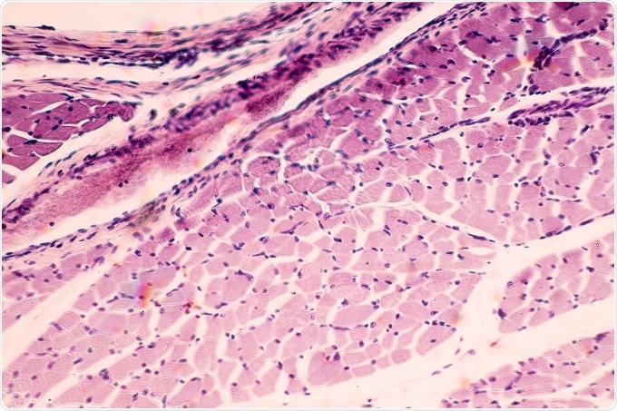
Cross section of Muscle Skeletal under the light microscope. ©Rattiya Thongdumhy / Shutterstock.com
The primary limitations of optical microscopes, including resolution, magnification and surface view, are described and discussed below. Additionally, some methods to overcome these limitations or alternative types of microscope that may be used instead are covered.
Resolution limit of optical microscopes
When an optical microscope with transmitted light is used at very high magnifications, the image of point objects may be distorted. They may be seen as fuzzy discs that are surrounded by diffraction rings, known as Airy discs.
These diffraction rings limit the ability of the optical microscope to resolve fine details of the sample. The resolving power of an optical microscope is a measure of the ability of the microscope to distinguish between two adjacent structural details, buy generic calcium carbonate nz without prescription without the interference of Airy discs.
The magnitude of diffraction and, hence, the resolving power of an optical microscope depends on the light wavelength (λ) and the numerical aperture (NA) of the objective lens. As a result, there is a distinct limit of an optical microscope to view adjacent structural details clearly, which is known as the diffraction limit of the microscope.
There are also several techniques that can be implemented to surpass the resolution limit of the transmitted light. For example, holographic techniques have been shown to reach a higher resolution in one experimental study. Other methods to increase the resolution of an optical microscope include:
- Spatially modulated illumination (SMI)
- Spectral precision distance microscopy (SPDM)
- Stimulated transmission emission depletion (STED)
- 3D super resolution microscopy
Additionally, with the use of a fluorescent sample, other techniques can also be used to reach a higher resolution. For example, near-field scanning optical microscopy with evanescent waves and stimulated emission depletion may prove useful.
Low magnification
The maximum magnification that can be achieved by an optical microscope typically ranges from 500x to 1500x. While this level of magnification has many purposes and can be useful for a number of practical applications, it is considerably lower than the magnification that can be achieved with electron microscopy. In contrast, an electron microscope may be able to provide magnifications greater than 160,000x.
As a result, the low magnification of an optical microscope is a limiting factor for some applications, when an electron microscope may be better suited to the purpose at hand.
Poor surface view
Similar to the magnification limitation of optical microscopy, the surface view of the sample with an optical microscope is sufficient for many purposes, but can be a limiting factor. It is significantly less clear than what can be achieved with an electron microscope, and this alternative may be preferred in some situations.
This does not limit its use for many applications though i.e. when high resolution with a clear surface view is not necessary. However, in some instances electron or another type of microscopy may be better suited to the purpose to achieve and maintain a high quality surface view at higher resolutions.
References
- http://www.nature.com/nprot/journal/v7/n9/fig_tab/nprot.2012.096_T1.html
- https://www.ncbi.nlm.nih.gov/pubmed/21668161
- https://www.ncbi.nlm.nih.gov/pmc/articles/PMC2645564/
- http://journals.aps.org/prl/abstract/10.1103/PhysRevLett.106.193905
- https://www.microscopyu.com/techniques/phase-contrast/specimen-contrast-in-optical-microscopy
Further Reading
- All Microscopy Content
- Advances in Fluorescence Microscopy
- Applications in Light Microscopy
- Electron Microscopy: An Overview
- Brief History of Microscopy
Last Updated: Aug 23, 2018
Written by
Yolanda Smith
Yolanda graduated with a Bachelor of Pharmacy at the University of South Australia and has experience working in both Australia and Italy. She is passionate about how medicine, diet and lifestyle affect our health and enjoys helping people understand this. In her spare time she loves to explore the world and learn about new cultures and languages.
Source: Read Full Article