vicodin 3601 is it strong
In a recent study published in Life Metabolism, researchers first used a diet-induced obese (DIO) murine model to test the effects of an extract of yellow petals of Dahlia pinnata as a novel treatment of type 2 diabetes (T2D).
Next, they performed a cross-over randomized controlled study in people with pre-diabetes and T2D to test its dose-dependent glucose-lowering effects, safety, and efficacy.
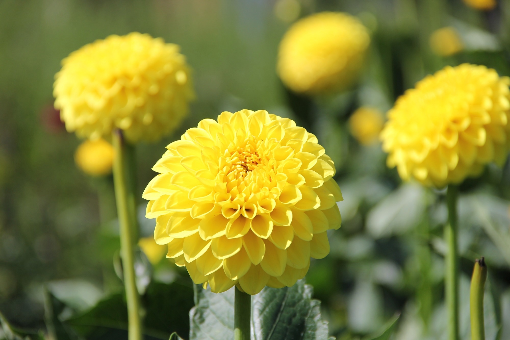
 Study: A dahlia flower extract has antidiabetic properties by improving insulin function in the brain. Image Credit: Shingo76Misumaru/Shutterstock.com
Background
Glucose intolerance and insulin resistance (IR) are key features of T2D pathogenesis. Claude Bernard discovered that the brain controls blood glucose levels back in 1885.
Around 150 years later, researchers revisited his findings and found that dysregulation of the central pathways of glucose homeostasis in the hypothalamus was considered the root cause of T2D.
Specifically, circulating insulin reaches the hypothalamus, binds its receptor, seroquel and ativan for sleep and autophosphorylates, which, in turn, recruits and phosphorylates insulin receptor substrate (IRS).
It activates the phosphatidylinositol 3-kinase (PI3K) – protein kinase B (AKT) pathway that initiates the cascade of metabolic effects of insulin.
Studies have shown that inflammation in the hypothalamus promotes IR via the nuclear factor kappa B kinase subunit beta (IKKβ)/nuclear factor kappa B (NF-κB) inflammatory pathway.
Thus, pharmacological inhibition of NF-κB in the arcuate nucleus (ARC) residing neurons of the hypothalamus could weaken glucose intolerance and help treat hypothalamic inflammation.
Preclinical studies have shown that IKKβ inhibitor butein markedly lowered glucose and sensitized insulin in DIO mice by targeting hypothalamic inflammation.
The bark of Toxicodendron vernicifluum, the Chinese lacquer tree, is the best natural source of butein, a rare flavonoid, albeit with limited medical use due to its toxicity.
About the study
In the present study, researchers tested the effect of D. pinnata's flower extract, a non-toxic ornamental flower plant containing butein.
They obtained 12 to 14-week-old male C57BL/6 mice and fed them a high-fat diet (HFD) to induce obesity. Mice fed with a low-fat diet (LFD) served as controls.
After four weeks, they administered one, 3.3, and 10mg/kg body weight (BW) dahlia extract by oral gavage to test mice. Likewise, controls received 0.9% sodium chloride (NaCl) containing 5% ethanol (EtOH). They also injected 1.5 g/kg BW glucose intraperitoneally (IP).
After one hour of dahlia extract administration, they performed an intraperitoneal glucose tolerance (ipGTT) test on all DIO mice. They used a glucometer to measure their blood glucose levels.
The team also tested the effects of other flavonoids, 10 mg/kg butein, sulfuretin, isoliquiritigenin, or their combinations in another cohort of mice.
After the IP administration of insulin, they also performed an insulin tolerance test (ITT) on a cohort of HFD or LFD-fed mice. Additionally, the researchers conducted chronic treatment studies in mice fed with HFD or the LFD ad libitum for five weeks.
Further, the team performed intracerebroventricular (ICV) infusion of PI3K inhibitors in mice.
These animals then received the dahlia extract (10 mg/kg body weight) by oral gavage one hour later, and after another 60 minutes, they were subjected to an ipGTT. The researchers also conducted immunohistochemistry (IHC) on mouse brain coronal cryosections.
Furthermore, the team studied the role of the IKKβ-NF-κB pathway using epifluorescence microscopy in Zebrafish incubated for six hours in HFD and five hours later in dahlia extract (2.75 µg/mL).
Finally, the team performed a clinical study among 13 males aged 18−70 with glycated hemoglobin (HbA1c) concentrations between 40−65 mmol/mol to test the effects of three doses of dahlia extract.
They first supplemented all participants with capsules containing 5, 20, or 50 mg of powdered dahlia extract.
Next, they subjected them to a baseline oGTT. Post-12-hour overnight fasting, they asked all the participants to drink 75 g of anhydrous glucose dissolved in water, drew their venous blood samples, and withdrew their blood samples every 30 min until three hours for glucose, C-Peptide, and insulin levels quantification.
The team compared the area under the curve (AUC) of the oGTT over three hours for the three doses of the dahlia extract to the AUC of the baseline oGTT. They monitored them for adverse reactions and tracked their complete blood count.
Results
In DIO mice, orally administered EtOH dahlia extract containing high amounts of butein improved glucose tolerance and insulin sensitivity.
These effects did not diminish after chronic treatment, suggesting it could help sustain glucose homeostasis in the long term. Moreover, chronic treatment did not alter the mice's liver morphology, liver fat content, or weight.
However, ICV application of butein in DIO mice was ineffective, reflecting the effect of leptin on intestinal barrier function and differences in blood-brain-barrier (BBB) function between HFD-fed and leptin-deficient mice.
The chalcone isoliquiritigenin and the aurone sulfuretin in combination with butein also elicited the glucose-lowering effects in DIO mice, likely by functionally interacting in vivo.
Further studies should investigate whether flavonoids present in the extract or their metabolites mediated the beneficial effects of the dahlia extract on glucose homeostasis.
At the molecular level, HFD feeding led to low-grade inflammation in the hypothalamus, ultimately leading to the development of T2D.
It also led to reactive astrogliosis in the hypothalamus, reflecting the pro-inflammatory nature of the HF diet, which contributed to the functional impairment of neuronal circuits governing energy homeostasis.
Hypothalamic insulin signaling, particularly PI3K, mediated the dahlia extract's glucoregulatory effects.
Further, the dahlia extract prevented the onset of astrogliosis after HFD feeding, and the improvement of glucose tolerance was associated with suppressing inflammatory signaling pathways within the hypothalamus.
In Zebrafish, the dahlia extract reduced hyperactivity of NF-κB pathway but did not decrease NF-κB activity to levels below controls.
Even though the dahlia extract improved glucoregulation in people with both pre-diabetes and T2D, in five participants with HbA1c ≥ 48 mmol/mol, the glucose-lowering effect of the 60 mg/m2 dahlia extract dose was more pronounced, suggesting dahlia extract was most effective in the treatment of patients who had already developed T2D.
Conclusions
To conclude, the current study results showed that the extract of yellow petals of D. pinnata restored glucose homeostasis in HFD-fed mice.
Furthermore, it was safe and effective in humans, necessitating further testing of its efficacy and safety as a therapeutic for people with T2D.
-
Pretz, D. et al. (2023) "A dahlia flower extract has antidiabetic properties by improving insulin function in the brain", Life Metabolism, 2(4). doi: 10.1093/lifemeta/load026. https://academic.oup.com/lifemeta/advance-article/doi/10.1093/lifemeta/load026/7200078?login=false
Posted in: Medical Science News | Medical Research News | Medical Condition News | Miscellaneous News
Tags: Blood, Brain, Chronic, Diabetes, Diet, Efficacy, Ethanol, Fasting, Flavonoid, Glucose, Glycated hemoglobin, HbA1c, Hemoglobin, Hyperactivity, Hypothalamus, IHC, Immunohistochemistry, in vivo, Inflammation, Insulin, Insulin Resistance, Kinase, Leptin, Liver, Metabolism, Metabolites, Microscopy, Morphology, Neurons, Obesity, Preclinical, Protein, Receptor, Type 2 Diabetes

Written by
Neha Mathur
Neha is a digital marketing professional based in Gurugram, India. She has a Master’s degree from the University of Rajasthan with a specialization in Biotechnology in 2008. She has experience in pre-clinical research as part of her research project in The Department of Toxicology at the prestigious Central Drug Research Institute (CDRI), Lucknow, India. She also holds a certification in C++ programming.