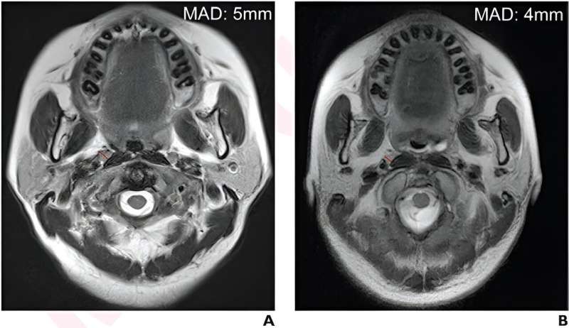
According to the American Journal of Roentgenology (AJR), using a 6-mm threshold, rather than a 5-mm threshold, helps facilitate better risk stratification and treatment decisions in patients with nasopharyngeal carcinoma (NPC).
“Future American Joint Committee on Cancer (AJCC) staging updates should consider incorporation of the 6-mm threshold for N-category and tumor-stage determinations,” wrote corresponding author Zhiying Liang, MD, from the radiology department at China’s Sun Yat-sen University Cancer Center.
This AJR accepted manuscript by Liang et al. included 1,752 patients (median age, 46 years; 1,297 men, 455 women) with NPC treated by intensity-modulated radiotherapy from January 2010 to March 2014 from two hospitals; 438 patients underwent MRI 3-4 months after treatment.
Two radiologists measured the minimal axial diameter (MAD) of the largest retropharyngeal lymph node (RLN) for each patient via consensus. Then, to assess interobserver agreement, a third radiologist measured MAD in 260 randomly selected patients. Initial ROC and restricted cubic spline analyses were used to derive an optimal MAD threshold for predicting progression-free survival (PFS).
Ultimately, in patients with NPC, overall survival was significantly different between patients with stage-I and stage-II disease defined using a 6-mm threshold (p = .04)—but not using a 5-mm threshold (p = .09). The 5-year PFS rate was associated with post-radiotherapy MAD ≥ 6 mm (HR = 1.68, p = .04) but not with post-radiotherapy MAD ≥ 5 mm (HR = 1.09, p = .71).
“Given the absence of a defined size threshold in the AJCC 8th edition staging manual,” the authors noted, “we propose that future updates to the manual incorporate this threshold for N-category and tumor-stage determinations.”
More information:
Yuliang Zhu et al, Optimal Size Threshold for MRI-Detected Retropharyngeal Lymph Nodes to Predict Outcomes in Nasopharyngeal Carcinoma: A Two-Center Study, American Journal of Roentgenology (2023). DOI: 10.2214/AJR.23.29984
Journal information:
American Journal of Roentgenology
Source: Read Full Article