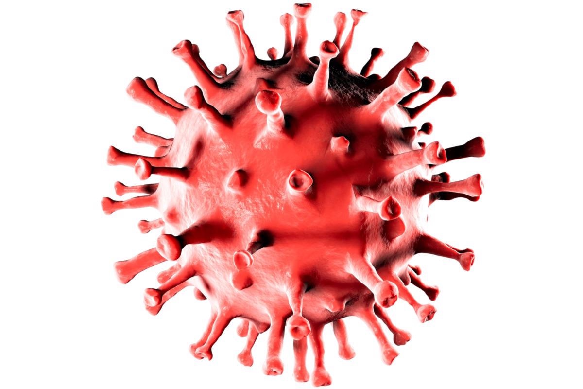
The severe acute respiratory syndrome coronavirus 2 (SARS-CoV-2), along with all other coronaviruses, possesses an organized nucleocapsid (N) protein that is formed by a ribonucleoprotein (RNP) complex and is surrounded by a lipid envelope.

Study: Structural dynamics of SARS-CoV-2 nucleocapsid protein induced by RNA binding. Image Credit: Naeblys / Shutterstock.com
Background
The N protein is one of the four structural proteins of coronaviruses and is the major component of RNP. Along with packaging the viral genome, the N protein is also required for viral replication, transcription, osteopathic school of medicine stratford nj translation, and assembly of the RNPs to viral particles.
The N protein consists of two structural and globular domains that can bind single-stranded ribonucleic acid (RNA) and DNA molecules, which are represented by the N-(NTD) and C-terminal (CTD) domains. Additionally, the N protein also consists of an extensive basic U-shaped RNA-binding cleft for the binding and melting of transcription regulatory sequences (TRS).
The CTD helps in protein dimerization and forms a positive groove that can recognize the packaging signal (PS), which leads to the assembly of the RNP into virion particles. Moreover, the flexible C-terminal tails also promote protein oligomerization.
The CTD and NTD are connected by an SR-linker, which has disordered serine and arginine-rich regions. Several studies have highlighted that the SR linker can be phosphorylated, which reduces the affinity of the protein for RNA and leads to liquid phase separation of the N protein with other viral proteins and RNA.
Recent studies have shown that the emergence of the highly transmissible SARS-CoV-2 B.1.1.7 strain is associated with SR-linker mutations. Therefore, understanding the orientation of NTD and CTD, as well as the contribution of the SR-linker and other unstructured regions to the overall N protein organization, is of importance. However, no three-dimensional (3D) structures of the full-length coronavirus N protein are currently available.
A new study published in the journal PLOS Computational Biology used electron microscopy, biophysical experimental analysis, molecular modeling, and molecular dynamics simulations to predict structure models of the full-length SARS-CoV-2 N protein in both the presence and absence of RNA.
About the study
The current study involved the isolation of SARS-CoV-2 RNA, reverse transcription to complementary DNA (cDNA), and cloning of the N protein sequence. The expression of the N protein was carried out in E. coli BL21 (DE3) cells and purified using size exclusion chromatography (SEC) and metal affinity.
The purified N protein was treated with RNase A or the purification was carried out in the presence of urea and high salt concentration for the production of N protein without any nucleic acid contamination.
Thereafter, protein samples were analyzed by FAR-UV circular dichroism, dynamic light scattering, size-exclusion chromatography multi-angle light scattering (SEC-MALS) analysis, negative staining and imaging, and image processing. Coarse-grained (CG) molecular dynamics simulations were performed, followed by a comparison with biophysical data. Finally, three-dimensional (3D) atomic model fitting was performed, along with all-atom molecular dynamics simulations.
Study findings
The 260/280 nm absorption ratio for the N protein treated with RNase A decreased from 1.8 to 0.6, while the absorption ratio for the urea treated N protein was 0.5. The circular dichroism profile for the urea treated and RNase A treated N protein was comparable.
Various rounded particles with dimensions less than 5 millimeters (mm) were detected in negative stain images of the urea-treated N samples. Analysis of these particles indicated that the area distributions of the urea-treated N protein fit well into the area distributions of the NTD monomer and CTD dimer. This suggests that these particles correspond to the globular domains of NTD and CTD.
The results of CG simulations do not indicate NTD-CTD contacts. Highly flexible conformations of full-length N protein are observed in the absence of RNA.
Moreover, a myriad of particles and aggregates were observed in N protein samples that were not treated with urea or RNase A. The larger aggregates were 20 to 30 nanometers (nm) in width, while the smaller aggregates were less than 20 nm in width. These particles were of dynamic dimensions and accordingly grouped into seven classes.
The N protein was also observed to undergo protein oligomerization and compaction in the presence of RNA, which was further confirmed by CG dynamics simulations. The all-atoms model suggested that the NTDs are oriented side-by-side facing the CTD dimer where all the domains help to shield the RNA segment. The arginine and lysine residues of the SR-linker were also found to interact with RNA and the serine residues remain exposed to solvents.
Furthermore, the 3D density map reconstruction indicated four quasi-globular units that are connected through the edges in a planar configuration where each globular unit contains an N dimer and the dimerization is brought about by van der Waals interactions between the C-terminal tail residues.
Conclusions
The current study highlights the higher-order oligomeric structure and dynamics of the SARS-CoV-2 N protein. This information can help to provide a better understanding of the multifunctional and versatile role of this protein.
- Ribeiro-Filho, H. V., Jara, G. E., Batista, F. A. H., et al. (2022). Structural dynamics of SARS-CoV-2 nucleocapsid protein induced by RNA binding. PLOS Computational Biology. doi:10.1371/journal.pcbi.1010121.
Posted in: Molecular & Structural Biology | Medical Science News | Medical Research News | Disease/Infection News
Tags: Arginine, Chromatography, Cloning, Contamination, Coronavirus, Coronavirus Disease COVID-19, DNA, Dynamic Light Scattering, E. coli, Electron, Electron Microscopy, Genome, Imaging, Lysine, Microscopy, Nucleic Acid, Protein, Respiratory, Ribonucleic Acid, RNA, SARS, SARS-CoV-2, Serine, Severe Acute Respiratory, Severe Acute Respiratory Syndrome, Syndrome, Transcription, Translation

Written by
Suchandrima Bhowmik
Suchandrima has a Bachelor of Science (B.Sc.) degree in Microbiology and a Master of Science (M.Sc.) degree in Microbiology from the University of Calcutta, India. The study of health and diseases was always very important to her. In addition to Microbiology, she also gained extensive knowledge in Biochemistry, Immunology, Medical Microbiology, Metabolism, and Biotechnology as part of her master's degree.
Source: Read Full Article