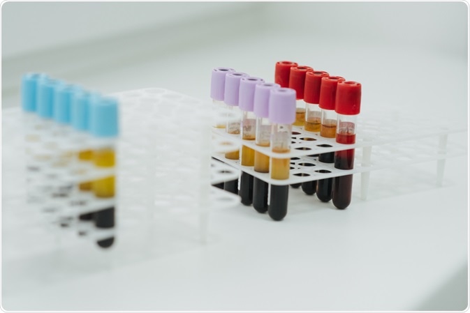
Researchers from the University of Texas have determined the procoagulant activity of clinical cellular therapeutics (CCTs). CCTs include human mesenchymal stromal cells (MSC) and mononuclear cells (MNC).
 Image Credits: Vitalii Vitleo / Shutterstock.com
The team determined that the acceleration of blood coagulation in trauma patients relative to healthy subjects is due to the MSC/MNC expression of tissue factor (TF). This effect was reversible with the anticoagulation drug heparin.
The study's findings build on the group's previous study in which blood samples from healthy donors received in vitro treatment with 33 CCT donors. In this current study samples from 36 trauma patients and 10 healthy controls were subject to in vitro treatment with 4 CCTs.
The principal aim of the present study was to illustrate CCT procoagulant effects in trauma patients; this is clinically relevant, as they represent a population which can clinically benefit from CCT.
Investigating the effect of CCT in trauma patients
Traumatic injury is associated with high levels of mobility as a result of associated conditions such as hemorrhaging. A potential solution for these morbid conditions is chemical cellular therapeutics which include MCS and MNCs; the application is limited due to the expression of tissue factor, a potent stimulant of coagulation.
TF expression is known to cause blood coagulation in non-trauma patients; the causal link between TF and coagulations has been demonstrated by using thromboelastography (TEG). TEG refers to the methodology of testing blood coagulation through the measurement of viscoelastic properties of whole blood clot formation under stress.
Despite this, the effects of MSCs or MNCs expressing TF in a population of patients with traumatic injury is uncertain. As such, the purpose of the group study was to determine the procoagulation activity of these CCTs in traumatic injury patient populations and its reversal.
The TEG methodology was used to measure blood clotting effects of CCT on the anticoagulant heparin was added to reverse this effect.
CCTs accelerate clot formation in trauma patients
A cohort of 36 trauma patients and healthy controls were selected to investigate the procoagulant effect of CCTs. The most prominent finding was a decrease in clotting time, referred to as R time, in response to an increasing TF load as the CCT treatment was increased. Notably, the R time was significantly faster in the trauma patient samples relative to healthy controls.
The addition of heparin resulted in the restoration of TEG clotting time to the control values for each tissue source of CCT.
The acceleration of TEG R time is associated with deep vein thrombosis, with trauma patients admitted with hypercoagulable TEG experiencing higher rates of DVT relative to those who do not. A study by Selby et al. found that soluble fibrin specifically relates to the initial formation of the fibrin clot.
Clinical considerations of CCT-associated TF
Consequently, the group demonstrated that CCTs increase the rate of clot formation in vitro. The procoagulant effects of these cells are an important consideration in the clinical setting and already suggested safety release criterion for clinical application. The team noted that cell sources with lower tissue factor or cell expansion methods that reduce the expression of TF are preferable alternatives.
Typically, traumatic injury results in a property of hypercoagulation relative to uninjured subjects. The hypercoagulable state experienced by trauma patients is thought to explain the greater procoagulant effect of CCT in healthy subjects.
The team noted that healthy subjects possess a greater time over which response to CCT treatment is possible because that baseline clotting time is longer. in comparison, trauma patients demonstrate accelerated clotting as a result of a natural reaction to trauma and so have a reduced capacity for additional procoagulant response.
Reversing the CCT procoagulant effect using heparin
Heparin exerts its effect by inactivating activated factor X. This limits, factor VII, the TF activation factor. TEG R time is restored to normal values for four different CCTs. The amount required was approximately double the amount to normalize the R time.
Compared to a previous study by Silachev et al, the group found that bolus administration used in this study is a more effective means of R time normalization relative to the systemic rate of heparin administration used by Silachev.
Issues of the procoagulant effect in severely injured trauma patient
Although the coadministration of heparin could mitigate the hypercoagulant properties of CCTs, in patients with traumatic brain injury heparin is contraindicated as bleeding can be exacerbated.
In addition, patients experiencing organ damage are susceptible to both bleeding and prophylactic anticoagulation. These instances represent cases in which Microthrombi deposition is likely and hence the procoagulant effect of CCTs fails to be an advantage.
Limitations of CCT-mediated TF in vitro
The team explored the procoagulant response to CCT in vitro; this limits the scope of the investigation as the TF exerts a systemic effect in vivo.
TF produces an envelope around blood vessels; under normal conditions, TF is not present in circulating blood cells and restricted to the adventitia. When CCTS are injected into the intravascular space, the TF released by these cells causes a rapid coagulation reaction or a blood-mediated inflammatory reaction.
The mechanism underpinning the consumption of coagulation factors mediated by TF released by the introduced CCTs, followed by the formation of fibrin monomers. This encourages what is known as consumptive coagulopathy, which accelerates the clotting process.
Once this consumptive coagulopathy is complete, the team hypothesizes that the TEG would indicate is slowing down of cutting, reduction in cloud formation and strength. The potential for this discrepancy to arise indicates the need to explore the in vivo effect of CCT-associated TF over time.
Source
George, J et al. (2020) Procoagulant in vitro effects of clinical cellular therapeutics in a severely injured trauma population. Stem cells Transl. Med. doi: 10.1002/sctm.19-0206.
Silachev, DN et al. (2019) Effect of MSCs and MSC‐derived extracellular vesicles on human blood coagulation. Cells. doi: 10.3390/cells8030258
Further Reading
- All Cellular Biology Content
- Structure and Function of the Cell Nucleus
- What Are Organelles?
- Ribosome Structure
- Protein Production: Initiation, Elongation and Termination
Last Updated: Mar 12, 2020

Written by
Hidaya Aliouche
Hidaya is a science communications enthusiast who has recently graduated and is embarking on a career in the science and medical copywriting. She has a B.Sc. in Biochemistry from The University of Manchester. She is passionate about writing and is particularly interested in microbiology, immunology, and biochemistry.
Source: Read Full Article