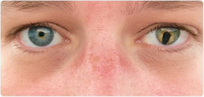
Ocular coloboma is an abnormality of the eye resulting from its defective development. One or both eyes may be affected by holes or gaps in the cornea, iris, ciliary body, lens, choroidal layer, fluoxetine hydrochloride as treatment lens, retina or optic disc. In many patients, a coloboma is accompanied by microphthalmia and anophthalmia, or other defects in various parts of the body.
Colobomas are present in 0.5–7.5 per 10,000 births, and can be caused by a genetic mutation or by toxic environmental factors.

What causes a coloboma?
As the baby develops in the womb, a specialized layer of ectoderm (the neuroectoderm), which gives rise to neural cells, comes to the surface to form the optic vesicle. This then invaginates, or curves inwards, to form two parts: the optic fissure in front and the optic cup towards the back. The optic fissure then closes as the two lips grow towards each other. Their fusion leaves only a small gap called the optic disc, through which the hyaloid artery enters the eye.
This fusion process occurs from the fifth to the seventh week of development. Any disruption can result in coloboma formation. The optic fissure is formed at the lower part of the eyeball, which means most colobomas are found there. The segment that remains unfused determines the part of the eye that is affected.
Some colobomas go unnoticed because they are very small and isolated. Others are larger but still isolated. More severe conditions result when a coloboma is associated with other abnormalities of the eye like microphthalmos. The most severe cases are when a coloboma is associated with anomalies of the brain and other systems outside the brain.
The classification of colobomas reflects the groups of coloboma genes involved and the period of development affected. Generally, the earlier in development a defect occurs, the more severe the congenital outcomes. Genes including SHH and SIX3 are involved in defects occurring before the 20th day of fetal development and result in severe anomalies of the eye, brain, and other organ systems. This is also traceable to the fact that SHH is a gene expressed in almost all tissues.
After this period, genes like TCOF1 may cause less severe defects of the brain and organs, whereas PAX6, MAF1, CHX10, or RBP4 can cause isolated colobomas. MAF1 and similar genes are expressed only in the eye; hence, their expression results in a milder phenotype.
Another factor that comes into play is the presence of genetic redundancy, which means that some gene anomalies can be compensated for by other genes expressed in certain tissues like the brain, but not in the eye, for instance, where the compensatory genes are not expressed.
Genetic causes
Most syndromes associated with coloboma are the result of Mendelian inheritance or chromosomal anomalies. As of now, almost 40 genetic regions linked to coloboma formation have been traced to their chromosomal origins, and many of the genes have been identified as well.
Some known coloboma-associated genes include SHH, CHX10 and MAF. Three coloboma syndromes share the same genetic locus at 22q11, making this a likely location for one or more genes that are crucial to normal development of the eyes.
Autosomal dominant and autosomal recessive inheritance are found equally in 27 coloboma phenotypes which are not yet mapped, whereas three are supposed to be X-linked. For some, the mode of inheritance is not yet clear. Thus colobomas are not caused by any one gene, but are rather a part of a more widespread aberration in development.
Environmental causes
Present research shows at least 39 genes are involved in coloboma syndromes, or isolated coloboma. All these are crucial to early intrauterine development, particularly of the central nervous system. However, these mutations account for only about half of all colobomas, which means many more mutations that could cause this condition remain unknown.
Most sporadic cases of coloboma are unilateral and often due to environmental factors, leading to malformations in multiple systems of the body. One classic example is the CHARGE syndrome, wherein colobomas of the iris or uvea is present in almost 86% of patients. The mechanism by which this occurs is not always clear, but such conditions comprise a good percentage of patients with coloboma.
Some suggested environmental triggers (operating during pregnancy) include:
- Drugs used in pregnancy, such as thalidomide (4%) and alcohol
- Vitamin A and vitamin E deficiency
- Infections with cytomegalovirus (CMV) and toxoplasmosis
- Ionizing radiation exposure
- Hyperthermia
Syndromes associated with coloboma
There are several conditions of which a coloboma forms a part, such as:
- Treacher-Collins syndrome
- Cat-eye syndrome where the coloboma is a vertical one in the iris, caused by an abnormality of chromosome 22q11
- Patau syndrome caused by an extra copy of chromosome 13 (trisomy 13)
- Coloboma with cryptophthalmos where the eyelids are absent
- CHARGE syndrome which stands for a constellation of Coloboma, Heart defects, Atresia of the nasal choanae, Growth retardation, Genital anomalies and Ear abnormalities, sometimes with microphthalmia
- Manitoba oculotrichoanal syndrome with multiple physical anomalies of the eyes, hair and anal opening
- Fraser syndrome with webbing and number abnormalities of the fingers or toes, or both, with renal and genital abnormalities and sometimes cryptophthalmos
- Goldenhar syndrome with faulty development of the ear, nose, soft palate and jaw
- Amniotic band syndrome
- Aicardi syndrome, Noonan syndrome, renal coloboma syndrome and Solomon syndrome
Sources
- https://www.ncbi.nlm.nih.gov/pmc/articles/PMC3257322/
- https://www.ncbi.nlm.nih.gov/books/NBK532877/#_article-21509_s6_
- https://ghr.nlm.nih.gov/condition/coloboma#genes
- www.ncbi.nlm.nih.gov/pmc/articles/PMC1735648/pdf/v041p00881.pdf
Further Reading
- All Coloboma Content
- What is Coloboma?
- What are the Symptoms Coloboma?
- Treating and Managing Colobomas
Last Updated: Mar 20, 2019

Written by
Dr. Liji Thomas
Dr. Liji Thomas is an OB-GYN, who graduated from the Government Medical College, University of Calicut, Kerala, in 2001. Liji practiced as a full-time consultant in obstetrics/gynecology in a private hospital for a few years following her graduation. She has counseled hundreds of patients facing issues from pregnancy-related problems and infertility, and has been in charge of over 2,000 deliveries, striving always to achieve a normal delivery rather than operative.
Source: Read Full Article