Neuroinflammation – a hallmark of neurodegenerative diseases, can be driven by a variety of triggers within the central nervous system (CNS). Such stimuli may include – pathogens, injury, toxic metabolites, and protein aggregates.
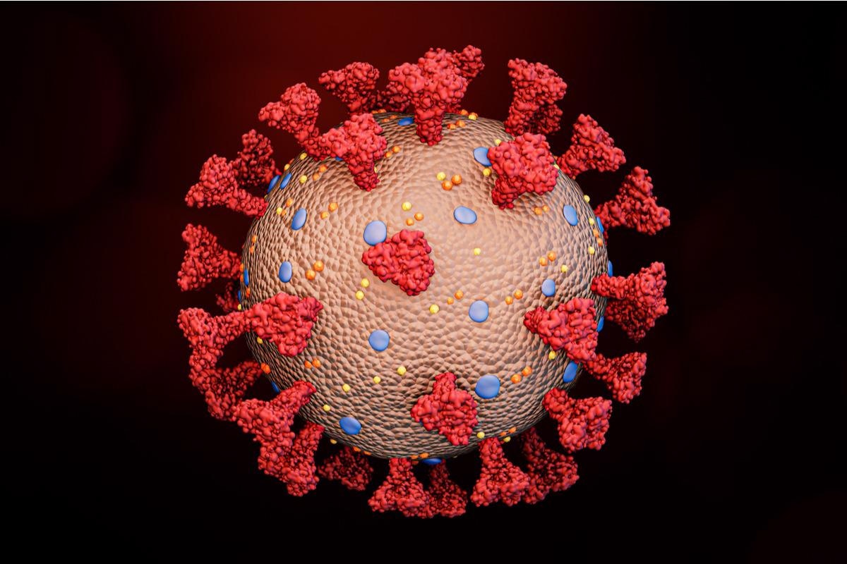
 Study: SARS-CoV-2 drives NLRP3 inflammasome activation in human microglia through spike-ACE2 receptor interaction. Image Credit: MattLphotography/Shutterstock
Among pathogens, viruses may also accelerate neurodegeneration, and this concept is gaining attention in the current coronavirus disease 2019 (COVID-19) pandemic. The severe acute respiratory syndrome coronavirus 2 (SARS-CoV-2) is known to invade the brain. Evidence suggests that SARS-CoV-2 patients exhibit signs of viral invasions in multiple organs, along with extensive microglial activation and pronounced neuroinflammation in the brainstem.
The study
The aim of a new study published in bioRxiv* preprint server was to assess the NLR family pyrin domain containing 3 (NLRP3) inflammasome activation in response to SARS-CoV-2 and its spike protein, and the effects of this exposure in the presence of a-synuclein protein aggregate fibrils.
Here, human monocyte-derived microglia (MDMi) cellular model was used, following an established protocol to obtain adult microglia.
Findings
The findings showed that SARS-CoV-2 isolates and even spike protein alone can prime and activate the NLRP3 inflammasome in human microglia through nuclear factor kappa B (NF-κB) and angiotensin-converting enzyme 2 (ACE2). Additionally, microglia exposed to SARS-CoV-2 or its spike protein potentiated a-synuclein mediated NLRP3 activation – indicating a possible mechanism for COVID-19 for precipitating movement disorders in some infected individuals.
The MDMi cellular model showed no secreted virus in the supernatant of infected MDMi and mouse primary microglia (mMi) cell culture supernatants. These contrasted with that in Vero E6 and Caco2 cells. Hence, microglia cells did not seem to allow SARS-CoV-2 replication in vitro.
MDMi expressed ACE2 mRNA, yet, at lower levels than that of Vero E6 and Caco2 cells. Western blot analysis revealed that ACE2 protein levels varied greatly in individual donors and elicited a differential pattern of expression than those of Vero E6 and Caco2 cells. The latter
The control cells (VeroE6 and Caco2) displayed an expected full-length size of the glycosylated ACE2 form of approximately 120 KDa, while microglia cells showed molecular weights of ~135 and ~100 kDa. Notably, similar patterns have been also found in endothelial cells and heart tissue from COVID-19 patients.
Viral binding to MDMi was evident. Higher levels of intracellular luciferase activity were found in microglia cells infected with the pseudo-virus compared to the non-glycoprotein control (NE). The results suggested that SARS-CoV-2 was capable of invading human microglia cells.
A high level of ZsGreen fluorescent protein expression was observed in Vero E6 cells whereas, no intracellular ZsGreen fluorescence was noted in MDMi. Thus, the virus could not establish replication in these cells. It was also noted that even in the absence of priming, SARS-CoV-2 exposure can directly activate the inflammasome in MDMi. In fact, the virus could both prime and activate the inflammasome.
In addition, even the spike protein was capable of both, priming and activating the inflammasome and could itself initiate inflammasome activation directly in human microglia. The spike protein was found to activate NLRP3 in human microglia-like cells through the ACE2 receptor. The vigorous virus-mediated inflammasome activation, in vivo, can be explained through the inflammasome activation by SARS-CoV-2 in MDMi without the need for priming. Furthermore, the viral spike protein could induce innate immune responses through NF-kB signaling.
Parkinsonism cases have been recorded after SARS-CoV-2 infection, which could have occurred due to an increased proinflammatory environment—precipitated by the blood-brain barrier (BBB) disruption, peripheral cell infiltration and microglial activation. Aging, poor health and ongoing synucleinopathies may enhance such complications, accelerate neuronal loss and increase the susceptibility to developing Parkinson’s disease (PD) post-SARS-CoV-2 infection.
Previous evidence has shown deterioration of motor performance and motor-related disability is seen in PD patients recovering from COVID-19. Animal studies have revealed Lewy body formation in the brains of SARS-CoV-2 infected macaques.
The results illustrate the molecular mechanisms of SARS-CoV-2 that cause the activation of microglia and lead to neurological manifestations. It was stated that spike protein-mediated priming and activation of microglia through the ACE2-NF499 kB axis may promote NLRP3 inflammasome activation leading to neuroinflammation and neurological phenotypes. However, this adverse effect could be amplified in the background of neurodegenerative disease, such as PD.
*Important notice
bioRxiv publishes preliminary scientific reports that are not peer-reviewed and, therefore, should not be regarded as conclusive, guide clinical practice/health-related behavior, or treated as established information.
- Albornoz, E., Amarilla, A., Modhiran, N., et al. (2022). SARS-CoV-2 drives NLRP3 inflammasome activation in human microglia through spike-ACE2 receptor interaction. bioRxiv. doi: 10.1101/2022.01.11.475947 https://www.biorxiv.org/content/10.1101/2022.01.11.475947v1
Posted in: Medical Research News | Medical Condition News | Disease/Infection News
Tags: ACE2, Aging, Angiotensin, Angiotensin-Converting Enzyme 2, Blood, Brain, Cell, Cell Culture, Central Nervous System, Coronavirus, Coronavirus Disease COVID-19, covid-19, Disability, Enzyme, Fluorescence, Fluorescent Protein, Glycoprotein, Heart, in vitro, in vivo, Inflammasome, Intracellular, Luciferase, Metabolites, Microglia, Monocyte, Movement disorders, Nervous System, Neurodegeneration, Neurodegenerative Disease, Neurodegenerative Diseases, Pandemic, Protein, Protein Expression, Receptor, Respiratory, SARS, SARS-CoV-2, Severe Acute Respiratory, Severe Acute Respiratory Syndrome, Spike Protein, Syndrome, Virus, Western Blot

Written by
Nidhi Saha
I am a medical content writer and editor. My interests lie in public health awareness and medical communication. I have worked as a clinical dentist and as a consultant research writer in an Indian medical publishing house. It is my constant endeavor is to update knowledge on newer treatment modalities relating to various medical fields. I have also aided in proofreading and publication of manuscripts in accredited medical journals. I like to sketch, read and listen to music in my leisure time.
Source: Read Full Article