Angiography involves the use of x-ray imaging to examine blood vessels. The images generated during an angiography procedure are known as angiograms.
First, a needle is placed into the femoral artery. All areas of the body can be addressed from this one site. After needle access is established, catheters and wires are threaded through the arterial system to the target area of interest.
When the imaging is performed, an iodine-based contrast medium is usually injected into the system. The medium highlights the blood moving through the vessels.
Angiograms are performed in hospitals with patients usually under local anesthesia. The procedure may take from 20 minutes to several hours, depending on the difficulty of the test and how much contrast is required.
The Need for Angiography
If a patient has problems with their circulation, a physician may suggest that the patient undergoes an angiography to determine what is causing the problems. The results of the test may also help the physician develop treatment options.
Angiography can be used to detect significant arterial disease, which can lead to such conditions as stroke, heart attack, gangrene, and organ failure. The imagery from coronary angiography can help physicians plan a patient’s treatment for angina and heart attacks.
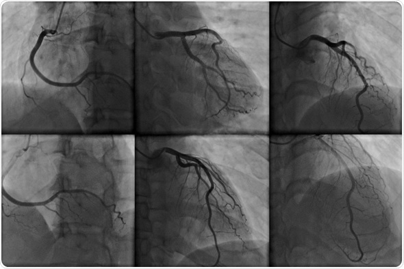
Types of Angiography
A variety of angiography procedures are available that can be harnessed to diagnose different medical conditions.
- Computed tomography angiography (CTA) utilizes x-rays, software, and hardware to produce horizontal, or axial, images, or slices, of blood vessels for diagnosis.
- Coronary angiography visualizes the inside of the coronary arteries. These images can locate stenoses in the arteries that may be responsible for chest pain, and which could cause a heart attack.
- Digital subtraction angiography (DSA) images blood vessels in the brain to check blood flow.
- Radionuclide angiography is a nuclear medicine procedure. A small amount of radionuclide (radiopharmaceutical or radioactive tracer), aids the examination of the target tissue. Resting radionuclide angiography assesses the heart's chambers in motion.
- Pulmonary angiography images the blood vessels to evaluate various conditions, such as aneurysm, stenosis, or blockages.
- Magnetic resonance angiography (MRA) uses magnetic resonance imaging (MRI) and contrast dye to visualize blood vessels. Physicians often use MRA to examine the heart and other soft tissues, and to assess blood flow.
- Renal angiography, also called arteriography, images the renal blood vessels to detect any signs of blockage or abnormalities affecting the blood supply to the kidneys.
Angiography Therapies
During an angiography, certain therapies can be performed, such as angioplasty or stent placement. Percutaneous coronary intervention (PCI), known as coronary angioplasty, is a non-surgical procedure that opens narrow or blocked coronary arteries.
The procedure restores blood flow to the heart muscle, which may have been blocked by plaque buildup. If the plaque ruptures, a blood clot can form on its surface.
A large clot has the potential to block the flow of blood through a coronary artery, a common cause of a heart attack. Over time, ruptured plaque also hardens and narrows the coronary arteries.
PCI can restore blood flow to the heart. During the procedure, a thin, flexible catheter with a balloon at its tip is threaded through a blood vessel to the affected artery, guided by x-ray imaging.
Once in place, the balloon is inflated to compress the plaque against the artery wall. This restores blood flow through the artery.
The procedure can optimize symptoms of coronary heart disease, including angina. The procedure also can lessen heart muscle damage caused by a heart attack.
Stents can be placed in arteries during PCI. Before the balloon is inflated, a stent is placed around it. When the tip of the catheter moves to the desired site, the balloon is inflated, pushing plaque against the artery wall. This widens the artery and helps restore blood flow. The fully extended balloon also expands the stent, pushing it into place in the artery.
The balloon is deflated and pulled out along with the catheter. The stent remains in the artery. Over time, cells grow to cover the mesh of the stent.
Potential Complications
With angiography, patients may experience bleeding or bruising where the artery was entered. They may have an allergic reaction to the contrast. Not often, the access artery may be blocked. Very rarely during angioplasty or stent placement, part of the arterial blockage can break off and travel to other arteries.
Sources
- http://www.nhs.uk/conditions/Angiography/Pages/Introduction.aspx
- www.nhs.uk/Conditions/Angiography/Pages/What-is-it-used-for.aspx
- stanfordhealthcare.org/…/radionuclide-angiogram.html
- https://vascular.org/patient-resources/vascular-tests/angiogram
- https://www.nhlbi.nih.gov/health/health-topics/topics/angioplasty
- http://www.nhlbi.nih.gov/health/health-topics/topics/stents/placed
Further Reading
- All Angiography Content
Last Updated: Feb 20, 2019

Written by
Joseph Constance
Joseph Constance has written about research, development, and markets in the health care and related fields. He has authored a number of articles, and business analysis/market research reports in the medical device, clinical diagnostics, and pharmaceutical areas. Joseph holds an MA from New York University in Communications. He enjoys spending time with his wife, biking, traveling, and learning about different cultures.
Source: Read Full Article