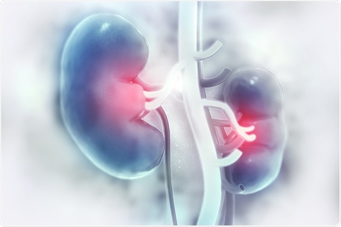
Medically known as ureterolithiasis, ureteral stone disease is characterized by the formation of calculi (i.e. stones) in the kidneys and obstructing the ureters. The ureters are the slender muscular tubes within our bodies that are responsible for the passage of urine from the kidneys to the urinary bladders.
Credit: crystal light/ Shutterstock.com
This flow is unidirectional if the anatomy and physiology of the urinary system are normal. In the Greek language, ureterolithiasis translates literally to “urine stone”. These stones may be of varying composition and produce different clinical features. This is because of varying signs and symptoms depending upon the site of impaction of the stones within the ureters.
Ureteral anatomy
The ureters are structurally composed of smooth muscle in the form of slender tubes, which are approximately 30cm in length in the adult. They arise from the proximally dilated part of each kidney, which is shaped like a funnel and is known as the renal pelvis. These tubes then wind down from each renal pelvis and enter the bladder on its posterior side. One tube enters on the right and the other on the left.
The junction formed between the ureter and the bladder is known as the ureterovesical junction, while that formed with the renal pelvis is known as the ureteropelvic junction. These two junctions, together with the point where the ureter crosses the bifurcation of the common iliac artery, constitute the 3 most common parts within the ureters where stones may become stuck. This is because they represent the sites at which the ureters are physiologically narrowed, thereby increasing the chances that a stone will lodge there during its downward course.
Clinical presentation
The main determinant of the signs and symptoms that are experienced by those affected is the anatomical location of the stone within the ureter. Furthermore, the degree to which the stone obstructs the passage of urine, as well as potential infections that may arise, both contribute to the clinical presentation.
Pain associated with ureteric calculi is typically abrupt in onset and is of a colicky nature, typically localized to the lower abdomen and flank, but may radiate to the pelvis and cause vomiting, nausea, and the appearance of blood in the urine. Stones that are impacted at the ureterovesical junction may give rise to increased frequency of urination that is accompanied with pain, while those at the ureteropelvic junction may cause pain that radiates to the lumbar region.
Calculi that are trapped at the entry of the ureters into the pelvis as it crosses over the bifurcation of the common iliac artery may cause anterior-inferior radiation of the pain. This can easily be confused with other abdominal pathologies. For instance, left-sided pain may mimic that seen in acute episodes of diverticulitis, while right-sided pain may resemble that of appendicitis.
References
- http://pmj.bmj.com/content/postgradmedj/27/305/128.full.pdf
- https://www.ncbi.nlm.nih.gov/pmc/articles/PMC2600100/
- https://radiopaedia.org/articles/ureter
Further Reading
- All Urolithiasis Content
- What is Urolithiasis?
- Causes of Ureteric Calculi
- What is Urolithiasis?
- Ureteric Calculi Prognosis
Last Updated: Feb 27, 2019

Written by
Dr. Damien Jonas Wilson
Dr. Damien Jonas Wilson is a medical doctor from St. Martin in the Carribean. He was awarded his Medical Degree (MD) from the University of Zagreb Teaching Hospital. His training in general medicine and surgery compliments his degree in biomolecular engineering (BASc.Eng.) from Utrecht, the Netherlands. During this degree, he completed a dissertation in the field of oncology at the Harvard Medical School/ Massachusetts General Hospital. Dr. Wilson currently works in the UK as a medical practitioner.
Source: Read Full Article