By Jeyashree Sundaram (MBA)
Malignant melanoma is a type of skin cancer that originates in the melanocytes. In the UK, about 15,000 individuals are diagnosed with melanoma annually, which means about 42 new cases crop up each day. In the past 10 years, the number of melanoma patients increased by about 50%, making this cancer the fifth most common among cancers in the UK.
Epidemiology
Women are more prone to develop the disease compared to men. Those at risk include individuals aged 65 years and above. Compared with African-Americans, white Americans are at a higher risk of developing melanoma. Whites with green or blue eyes, blond or red hair, and fair skin (which burns easily in the sun) are at a higher risk.
The risk factors include exposure to ultraviolet rays, multiple moles, a family history of melanoma, a personal history of skin cancer or melanoma, a weakened immune system, and the rare condition xeroderma pigmentosum.
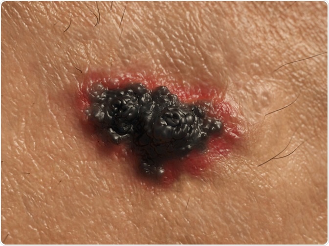
Disease Types
Although melanomas can develop over any part of the skin, the areas most commonly affected include the legs (in women), the chest and back (in men), the face and the neck. It affects areas such as the eyes, genitals, anal area, and mouth less frequently.
In superficial spreading melanomas, the disease grows outwards and not deeper into the skin. This pattern of growth means that the disease may not spread to other body parts. In nodular melanoma, the disease grows deep into the skin, often manifesting as a raised area on the surface of the skin, with a black or dark brownish color.
Lentigo maligna is a type of melanoma that develops from lentigo maligna of the skin. The disease grows deeper into the skin layers and may create nodules. As this type occurs due to excessive exposure to the sun, individuals who spend most of their time outdoors are frequently affected; this melanoma is most commonly seen on the face.
Acral lentiginous melanoma is seen on the soles and palms. It is most frequently found on the feet, around the area of the big toenail, and grows further under the nails. This rare type is seen most often in people with dark skin.
When the melanoma cells make melanin, the color of the melanoma tumors is black or brown. In case the melanoma cells do not produce melanin, the tumors may be pink, white, or tan in color.
Amelanotic melanoma types are rare and are usually colorless; they may also have a tinge of pink or red, making them difficult to diagnose, and being mistaken for other skin conditions.
Diagnosis and Staging
Although less common compared to squamous cell skin cancers and basal cell cancers, malignant melanomas can be very dangerous; when not diagnosed early, the disease can spread across the body.
Dermatologists ask the patients about the mole, such as for how long it has been there and if it has undergone any changes since its origin. They may apply oil on the affected skin and use a dermatoscope to magnify and examine the mole. They also examine the rest of the skin, and will take photographs of the mole and surrounding area at each visit, to compare and check for any changes that have been happening over time.
The Clark scale is used to stage melanoma. It is based upon the depth and thickness of the cancer cells that have spread into the layers of skin.
There are five levels in the Clark scale:
- Level 1 is when the melanoma cells are seen in the epidermis
- Level 2 is when the melanoma cells are present in the papillary dermis
- When the melanoma cells extend throughout the papillary dermis and into the reticular dermis, i.e., the next layer, it is termed Level 3
- When the melanoma cells have spread deep into the dermis, it is defined as Level 4.
- Level 5 is where the melanoma cells have penetrated into the subcutaneous fat layer
The Breslow scale depends upon the measured thickness (in millimeters) of the extent of melanoma cells within the skin surface. It is used in the TNM (tumor, node, and metastasis) staging of melanoma.
Treatment and Prevention
After the dermatologists confirm the diagnosis of malignant melanoma by an excision biopsy, a second operation is performed to remove a wide margin of skin. However, if the mole is likely to be benign but not unequivocally declared to be so, it is monitored for about 3 months before suggesting a course of action.
Although gender, family history, and race are risk factors that are not modifiable, there are some ways to reduce the risk of developing the disease:
- Limiting skin exposure to ultraviolet rays
- Watching the skin for the emergence of abnormalities in an existing mole, unusual moles, or new moles
- Maintaining a healthy immune system
References
- http://about-cancer.cancerresearchuk.org/about-cancer/melanoma/stages-types/clark-breslow-staging
- http://about-cancer.cancerresearchuk.org/about-cancer/melanoma/getting-diagnosed/tests-diagnose/looking-your-mole-dermoscopy
- http://about-cancer.cancerresearchuk.org/about-cancer/melanoma/treatment/surgery/surgery-remove-melanoma
- http://about-cancer.cancerresearchuk.org/about-cancer/melanoma/stages-types/types
- https://www.cancer.org/cancer/melanoma-skin-cancer/causes-risks-prevention/risk-factors.html
- http://about-cancer.cancerresearchuk.org/about-cancer/melanoma/about
- https://www.cancer.org/cancer/melanoma-skin-cancer/about/what-is-melanoma.html
- https://www.cancer.org/cancer/melanoma-skin-cancer/causes-risks- prevention/prevention.html
Further Reading
- All Melanoma Content
- What are Melanomas?
- Melanoma Symptoms and Cause
- How is Melanoma Diagnosed and Treated?
- Melanoma Prognosis
Last Updated: Feb 27, 2019
Source: Read Full Article