Increasingly, tattoos are becoming more commonplace; however, this trend has raised safety concerns. There are toxicological and biokinetic considerations to be made in regard to tattoos. However, animal experiments that are necessary to address these safety concerns are considered to be unethical, as, like cosmetics, they lack medical necessity.

Tattooing. Image Credit: Olena Yakobchuk/Shutterstock.com
Among these safety concerns are those related to the immune system. This is because tattooing involves the insertion of ink into tiny punctures created in the epidermis, and pigments are considered to be a foreign body that may elicit the immune response.
How does tattooing work?
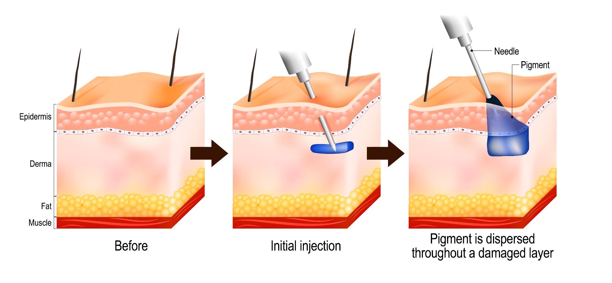
Tattoos are permanent depositions of insoluble pigment into the dermal skin layer. After tattoo inks are injected into the skin, ink particles may either be passively transported via the blood and lymph fluids, or subject to phagocytosis by immune cells, after which they are deposited in lymph nodes. After healing is complete, the dermis contains particles – as do the sinusoids of the draining lymph nodes.
What does tattoo pigment consist of?
Tattoo pigments consist of either inorganic metals and their oxides, or polyaromatic compounds; all of these are considered to be biologically inert. These metals include nickel, chromium, manganese, and cobalt. Alongside carbon black, the most used ingredient is titanium dioxide. This is a white pigment that is used to create shades when mixed with other colored compounds. The toxicity of titanium dioxide is dependent on its crystal structure, the photo catalytically active anatase structure, which can produce reactive oxygen species when exposed to daylight.
According to a paper published in 2017 in the journal Scientific Reports, there is analytical evidence to support the transport of organic and inorganic pigments, alongside toxic element impurities From tattoo ink – which eventually reach the lymph nodes. Scientists from the European Synchrotron Radiation Facility (ESRF) in Grenoble, France, conducted X-ray fluorescence measurements to identify the locations of titanium dioxide in both the micro and nano range in the skin and lymphatic system.
The researchers observed the transport of organic pigments, heavy metals, and titanium dioxide from the site of injection to regional lymph nodes. Organic pigments displayed the largest range in size with the smallest (nano) particles reaching the lymph nodes.
In addition to the changes in migration, the researchers were able to detect changes in the structure of the tissues adjacent to the tattoo particles. This was mediated through altered ratios of amide I α-helix to β-sheet protein and increased lipid contents.
Taken together, the authors reported strong evidence for both migration and long-term deposition of tattoo pigment, alongside conformational alteration of biomolecules which may cause cutaneous inflammation and other adverse events because of tattooing.
Tattoos and the immune response – Maintaining tattoos long-term
The dermis layer of the skin, the site at which tattoo ink is deposited, is rich in blood vessels, lymphatic vessels, nerves, and other structures. In the dermis, there are dermal dendritic cells, macrophages, CD4+ T cells, and innate lymphoid cells. These cells are well-placed to react in response to damage, such as that caused by a tattoo needle that enters the skin to inject pigment. The ink pigment is regarded as a foreign body, which elicits an immune response in an attempt to clear it.
Newly tattooed skin swells, and while the majority remains at the site of deposition in the dermis, regional lymph node migration is observed as discussed above.
When tattoo ink persists in the skin, the dermal macrophages phagocytose the ink. The ink is sequestered in their vacuoles. The enzymes present in the vacuole are not able to decompose the ink. According to a study conducted in 2018, researchers determined the origin, identity, and dynamics of the skin myeloid cells that are responsible for capturing and retaining these tattoo pigment particles.
These researchers demonstrated that these myeloid cells are exclusively comprised of dermal macrophages and not resident dermal cells. That is, other cell types play no role in containing the pigment that is injected into the dermal layer. Furthermore, deletional analysis demonstrated that the tattoo pigment particles are capable of undergoing cycles of capture-release-recapture without the risk of losing the tattoo at the site of injection.
The researchers used diphtheria toxin to selectively delete cells expressing CD64, a monocytic marker. CD64 is expressed on monocytes, macrophages, precursors to myeloid cells, and follicular dendritic cells. Through the generation of mice that were engineered to have the diphtheria receptor under the control of the CD64 gene, all myeloid cells were specifically targeted for ablation (i.e., deletion).
The researchers found that macrophage death results in the release of the trapped pigment; however two days following treatment with diphtheria toxin, the macrophage pool is replenished by circulating macrophages, which in turn, have been replenished by bone marrow-derived monocytes. These nearly derived macrophages subsequently engulf the free ink released by the prior macrophage population – and this process continues indefinitely which ensures the permanence of tattoos.
Therefore the researchers concluded that the persistence of tattoos long-term is dependent on the macrophage renewal rather than macrophage longevity.
 Process of tattooing. Image Credit: Designua/Shutterstock.com
The role of tattoos in immune-boosting
A study published in 2016 measured the immune function using secretory immunoglobulin A (SIgA) and cortisol present in the saliva of 29 participants to investigate the ‘inoculation hypothesis’ of tattooing. This inoculation hypothesis suggests that tattooing inoculates the immune system, inducing increased protection against stressors that produce soft tissue damage.
The tattoo experience was measured as a sum of the number of tattoos, lifetime hours tattooed, the number of years since the participants' first tattoo, the percentage of the body covered, and the number of tattoo sessions.
Researchers observed an inverse relationship between SIgA and cortisol, with less immunosuppression of SIgA observed in those who had more tattoo experience. Those that had no pre-existing tattoos experienced a greater strain on their immune system, observed by a larger dip in their SIgA.
Taken together, the data suggest that the body habituates to the external insult of tattooing over time. It is important to note, however, that the authors noted that individuals with healthy immune systems heal faster, increasing their likelihood of subsequent tattooing experience.
References
- Baranska A, Shawket A, Jouve M, et al. Unveiling skin macrophage dynamics explains both tattoo persistence and strenuous removal. J Exp Med. 2018;215(4):1115-1133. doi:10.1084/jem.20171608.
- Lynn CD, Dominguez JT, DeCaro JA. Tattooing to "Toughen up": Tattoo experience and secretory immunoglobulin A. Am J Hum Biol. 2016;28(5):603-9. doi: 10.1002/ajhb.22847.
- Schreiver I, Hesse B, Seim C, et al. Synchrotron-based ν-XRF mapping and μ-FTIR microscopy enable to look into the fate and effects of tattoo pigments in human skin. Sci Rep. 2017;7(1):11395. doi:10.1038/s41598-017-11721-z
Further Reading
- All Tattoos Content
- The Benefits and Risks of Laser Tattoo Removal
- Are Tattoos Safe?
Last Updated: Apr 27, 2022

Written by
Hidaya Aliouche
Hidaya is a science communications enthusiast who has recently graduated and is embarking on a career in the science and medical copywriting. She has a B.Sc. in Biochemistry from The University of Manchester. She is passionate about writing and is particularly interested in microbiology, immunology, and biochemistry.
Source: Read Full Article