Both acute and chronic stress in many different forms can lead to a condition known as acute thymic atrophy, which can severely impact the health of the immune system.
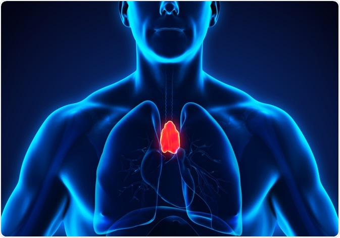
Thymus. Image Credit: Nerthuz/Shutterstock.com
What is the thymus?
Except for jawless fish, the thymus is found in all vertebrates. The thymus is a primary lymphoid organ located above the heart that plays a crucial role in the ability of the immune system to fight against pathogens, tumors, antigens, and other mediators of tissue damage. The immune system is often subdivided into either thymus-independent or thymus-dependent arms.
Thymus-independent immune system
The thymus-independent components of the immune system are otherwise commonly referred to as the innate immune system. The innate immune system is the body’s first line of defense against pathogenic invasion, which is achieved through macrophages, neutrophils, dendritic cells (DCs), and granulocytes.
These innate immune cells are activated by germline-encoded pattern recognition receptors, some of which include Toll-like receptors and nucleotide oligomerization domain (NOD)-like receptors. Once activated by these receptors, the macrophages and neutrophils will eliminate antigens through phagocytosis. Comparatively, DCs and other innate immune cells will process antigens and eventually serve as antigen-presenting cells for the adaptive immune system.
Thymus-dependent immune system
The thymus-dependent immune system, which is otherwise known as adaptive immunity, is achieved through the actions of both T lymphocytes and B lymphocytes (T and B cells, respectively). T and B cells, which both originate from the bone marrow, express specific antigen recognition receptors and are capable of developing highly specialized effector functions to create long-lasting immunologic memory.
Whereas B cells will be exported to the periphery after leaving the bone marrow, the development, maturation, and export of T cells all take place in the thymus. Through thymopoiesis, the thymus will utilize several different processes to ensure that the T cells are capable of recognizing true antigens, rather than inducing autoimmune reactivity.
For example, the thymus conducts a negative selection process that eliminates potentially self-reactive T cells and releases T cells that can recognize antigens into the periphery.
Stress-induced thymic atrophy
Various physiologic conditions including malnutrition, emotional distress, pregnancy, or pathologic conditions like infection, disease, cancer treatments, or regimens used to prepare patients for bone marrow transplants can cause stress on the body.
Regardless of the cause, stress disrupts the homeostatic balance of the immune system and can lead to a condition known as acute thymic involution. Some of the hallmarks of this thymic response to stress include a reduction in double-positive (DP) thymocytes and reduced output of naïve T cells to the periphery; both of which can significantly reduce the size of the thymus gland.
Infectious causes of thymic atrophy
Viral, bacterial, and fungal infections are common causes of acute thymic atrophy. Of particular concern is the human immunodeficiency virus (HIV), which can cause an infection that severely alters the normal function of the thymus as a result of the loss of developing thymocytes and thymic stromal cells.
In addition to HIV/AIDS, acute sepsis, which is a bacterial infection that is often acquired after surgery, can also cause acute thymic atrophy. To this end, sepsis can result in massive lymphocyte apoptosis which leads to impaired T and B cell responses and, ultimately, severe thymic atrophy.
Environmental stressors
Several different environmental stressors have been found to have a negative impact on human thymopoiesis and ultimately reduce thymus function until the stressor has been removed. Some examples of these stressors include malnutrition, starvation, alcoholism, and prolonged physical and/or emotional stress.
Stress due to the abuse and neglect of children, for example, has been found to lead to thymic involution that can significantly reduce the size of the child’s thymus. In fact, one autopsy of an eight-year-old child who was tortured by his family for several years found that the boy’s thymus weighed about 10 grams. When compared to a normal eight-year-old boy who should have a thymus gland weighing about 100 grams, this level of thymic atrophy was astounding.
Clinical situations and stress-induced thymic atrophy
Unfortunately, several pharmacotherapies are associated with thymic atrophy. Chemotherapy and irradiation therapy, both of which are used in the treatment of cancer patients, as well as cyclosporine, which is a drug that is widely used for the treatment of autoimmune diseases, have been associated with reduced thymus function and delayed immune reconstitution. The preparative regimens used before a bone marrow transplant are also associated with these adverse effects on the thymus.
Several studies have found that patients who have undergone cancer treatments or bone marrow transplant preparation protocols exhibit significantly reduced thymus function that is accompanied by peripheral T cell compartment with reconstitution. However, young patients who have previously undergone cancer treatment appear to recover from this reduced thymus function.
To this end, several pediatric cases have shown that after their treatment has been ceased, these patients exhibit an increase in CD45RA+ cells, extensive peripheral T cell reconstitution, and, ultimately, an increase in the size of their thymus.
Although this recovery has been identified there is a delay between the cessation of chemotherapy and the point at which the patient’s thymic function improves. Therefore, the patient is particularly vulnerable during this period to opportunistic infections.
References:
- Thapa, P., & Farber, D. L. (2019). The Role of the Thymus in the Immune Response. Thoracic Surgery Clinics 29(2); 123-131. doi:10.1016/j.thorsurg.2018.12.001.
- Gruver, A. L., & Sempowski, G. D. (2008). Cytokines, leptin, and stress-induced thymic atrophy. Journal of Leukocyte Biology 84(4); 915-923. doi:10.1189/jlb.0108025.
- Doctor describes injuries to Gabriel Fernandez’s body, testifies he was determined brain dead [Online]. Available from: https://abc7.com/palmdale-boy-child-abuse-gabriel-fernandez-torture-isauro-aguirre/2572619/.
- Tanegashima, A., Tamamoto, H., Yada, I., et al. (1999). Estimation of stress in child neglect from thymic involution. Forensic Science International 101(1); 55-63. doi:10.1016/S0379-0738(99)00003-1.
Further Reading
- All Stress Content
- Why can Uncertainty Cause Stress?
- Stress and Alzheimer’s Disease
Last Updated: Aug 23, 2021

Written by
Benedette Cuffari
After completing her Bachelor of Science in Toxicology with two minors in Spanish and Chemistry in 2016, Benedette continued her studies to complete her Master of Science in Toxicology in May of 2018.During graduate school, Benedette investigated the dermatotoxicity of mechlorethamine and bendamustine; two nitrogen mustard alkylating agents that are used in anticancer therapy.
Source: Read Full Article