Single-particle cryogenic electron microscopy (cryo-EM) is an analytical technique used to deduce the 3D structure of macromolecules (particularly proteins) as an alternative to X-Ray crystallography. It is not commonly used in quotidian practice but does offer some advantages over other techniques.
Skip to:
- How does single-particle cryo-EM work?
- What makes cryo-EM special?
- Using single-particle cryo-EM for protein analysis
- Advantages
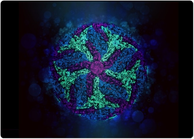 molekuul_be | Shutterstock
How does single-particle cryo-EM work?
Pure protein is applied to a grid with very small holes, normally supported by a metal framework. This will ideally evenly distribute the protein molecules throughout these holes in the grid, in varying orientations.
This grid is then snap frozen by immersing in a solution like liquid nitrogen or dry ice. This initially creates a stable snapshot of the protein in various orientations, and also prevents some damage to the protein, as well as averts the evaporation of the buffer.
Images are taken of all the individual proteins in this grid, and then aligned and averaged to create a 3D map. Lots of processing and analysis goes into the final steps of this data, proscar idaho finally producing a 3D model of the protein structure.
What makes cryo-EM special?
Single particle cryo-EM creates a more distinct snapshot of the behavior of a protein at a certain point in time, compared to crystallography where proteins have to form an ordered lattice together before the structure can be determined.
Due to this difference, cryo-EM can potentially give a more accurate insight into the natural form of a protein and provide more information about how it may function.
Allowing the protein to be free-moving up until a snapshot is taken can be a challenge though, as it makes the computational analysis of a protein much more difficult, because many images of the protein in different positions and orientations have to be collated together.
Using single-particle cryo-EM for protein analysis
The determination of the 3D structure of proteins represents the main application of this technique. The other two main methods of deducing protein structure is X-Ray crystallography or nuclear magnetic resonance (NMR).
The process of crystallography can be very unreliable and lengthy. The exact conditions of the protein’s environment have to be found in order for the protein to form a regular lattice. Many screens are, therefore, needed to produce the best quality crystals in order to produce X-Ray diffraction data.
Relatively large amounts of samples are needed for crystallography and NMR compared to single particle cryo-EM. This is a big advantage of cryo-EM, meaning that it can potentially be easier and cheaper in comparison to other methodologies.
Single particle cryo-EM can also be used for all sizes of protein, while NMR has a size restriction for the proteins that can be analyzed. This gives cryo-EM a larger range of options for protein structure analysis.
Advantages
Due to the simple and reliable methodology of single-particle cryo-EM, it is possible to analyze not only simple proteins, but also protein complexes and polymers like F-actin and microtubules, as well as transcription initiation complexes. Many protein complexes only exist for very short amounts of time, and this technique allows them to be visualized.
Other complexes that cryo-EM has enabled close study of are nuclear complexes, where protein binds to DNA or RNA. Many important aspects of cellular machinery rely on these complexes, including during DNA replication and RNA synthesis.
There are many structures in which crystallography cannot determine the structure of, yet single particle cryo-EM can. One example of these are membrane proteins. The transmembrane domain of these proteins needs to be stabilized in solutions by detergents, which can make the crystallization of proteins rather difficult.
Cryo-EM can also determine the structure of proteins with flexible regions without having to remove them, which is often done in crystallography. This cuts out a potentially very difficult step.
Sources
- Doerr, A. (2016). Single-particle cryo-electron microscopy. Nature Methods. nature.com/articles/nmeth.3700.
- Cheng, Y., et al. (2015). A Primer to Single-Particle Cryo-Electron Microscopy. Cell. doi.org/10.1016/j.cell.2015.03.050
- Nogales, E. (2016). The development of cryo-EM into a mainstream structural biology technique. Nature Methods. nature.com/articles/nmeth.3694
- Fernandez-Leiro, R. & Scheres, S. H. W. (2016). Unravelling biological macromolecules with cryo-electron microscopy. Nature. nature.com/articles/nature19948
Further Reading
- All Electron Microscopy Content
- Electron Microscopy: An Overview
- Applications of Electron Microscopy
- Advantages and Disadvantages of Electron Microscopy
- What is Transmission Electron Microscopy?
Last Updated: May 2, 2019
Written by
Jack Davis
Jack is a freelance scientific writer with research experience in molecular biology, genetics, human anatomy and physiology, and advanced analytical chemistry. He is also highly knowledgeable about DNA technology, drug analysis, human disease, and biotechnology.
Source: Read Full Article