An Overview of CMOS
The Healthcare Burden
Imaging Cells
Future Prospects
References
An Overview of CMOS
Complementary Metal Oxide Semiconductor (CMOS) is a type of circuit known to be cost-effective and efficient. These circuits are found in many day-to-day items, such as batteries and the image sensors components of digital cameras.
The biggest advantage of using CMOS components is that they are incredibly efficient. They don't need an input of electrical current unless they are changing between states, and as the semiconductors cooperate and limit the voltage output when not needed, they use minimal power and give off less heat than other designs.
The CMOS circuits consist of their sensory elements coupled with an array of circuitry necessary for an array of basic functions such as amplifying, filtering, and controlling the signal, as well as analog-to-digital conversion. The current technology can produce circuits as small as 0.1µm, although the current research works to produce them as small as 22nm.
These miniaturized circuits have many applications, although the most important factor to consider when using these circuits in a diagnostic setting is increased sensitivity. Although miniaturization and low power consumption would increase the product's portability, perusing these things shouldn't sacrifice sensitivity.
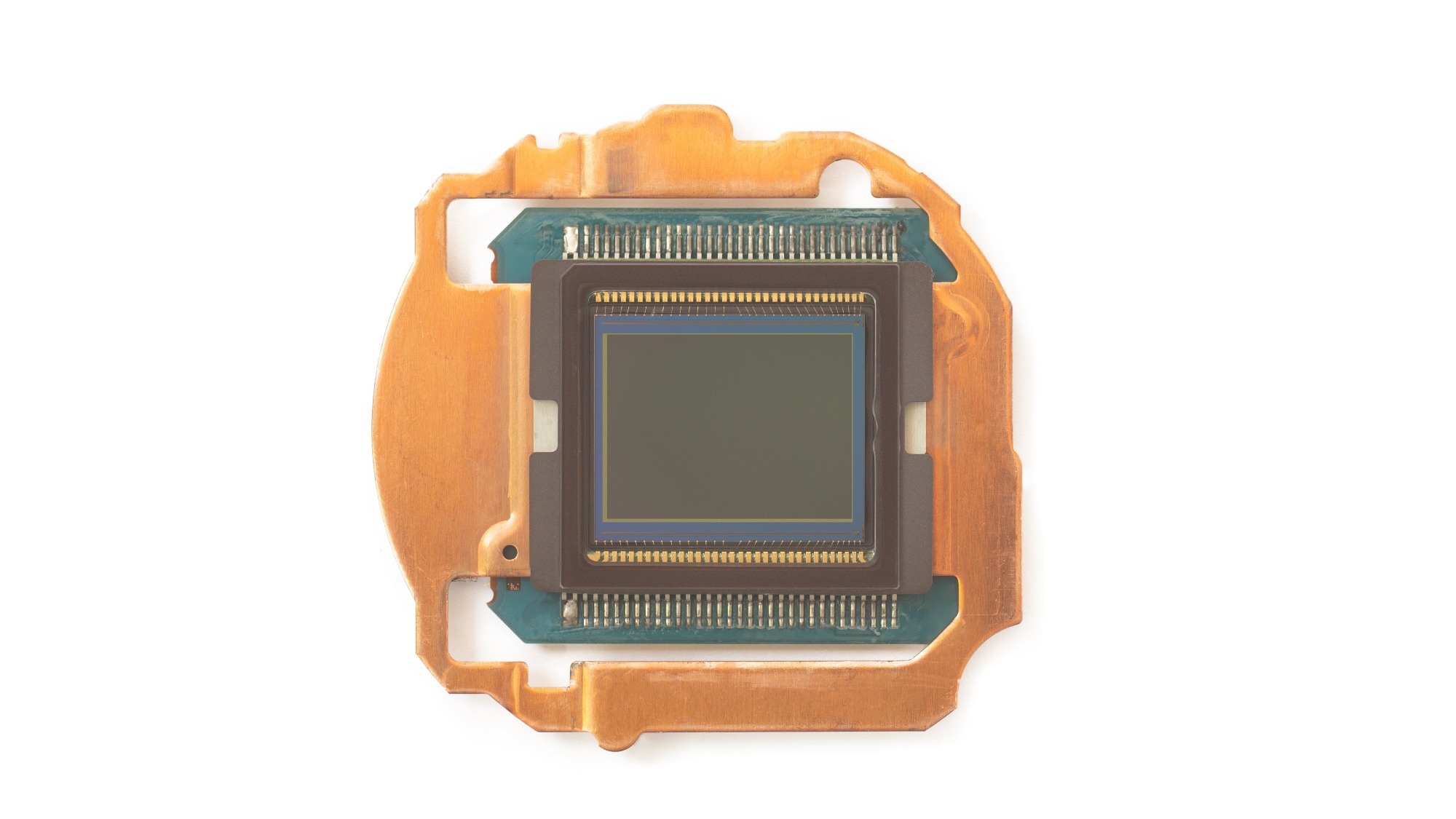
Image Credit: Teerasak Ladnongkhun/Shutterstock.com
The Healthcare Burden
The demand for reliable and cost-effective healthcare services is growing fast due to the increasing population size and age. The unfair distribution of wealth and climate change also increase the pressure on the healthcare system. These pressures have increased the importance of effective, cheap, and sensitive tests for a range of conditions.
CMOS-based products fill these criteria and so are in high demand. As these systems have already been tested and established, as well as being portable and having low-power usage, they have the potential to become essential in studying cells and diagnosing their conditions.
Antibodies eBook

Other cell counting methods, such as fluorescents imaging, require the cells to be tagged and more set up than when using CMOS devices. It is also not as efficient or accurate when the cells are counted manually using a microscope, which is the most viable option if there is no access to an algorithm.
Automated counting of cells is usually done using an algorithm that can include a CMOS system. If a viable cell count is needed, then CMOS systems provide a reliable alternative to the conventional patch-clamp technique and provide non-invasive real-time results on living cells.
Conventional techniques for observing cells require cell culturing facilities and optical and spectroscopic equipment. New technologies are making cheaper, more effective options more accessible and providing various options depending on the specific situation. CMOS systems provide a cheap, portable, and efficient alternative to the established methods.
Imaging Cells
Unlike conventional techniques, capacitive biosensors which utilize CMOS systems do not require the cells of interest to be fluorescently tagged. This provides a more accurate result as it is thought that the cell parameters could be affected in preparing the cells for fluorescent tagging.
The capacitive biosensors detect the charge sharing between cells and translate this to output voltages that can be detected and interpreted. The growth medium in which the cells are grown, the supporting substrate, and the cell parameters all affect the output voltage detected.
When subjected to low electrical fields, ionic clouds surrounding the cell polarizes, generating the cell's capacitance behavior. The capacitance of healthy cells is higher than that of damaged or dead cells, and they adhere to surfaces more readily. Detecting the capacitance of cells is a viable alternative to the tedious task of cell counting.
Common problems in imaging and identifying cells are the efficient separation of specific cells from whole blood and the rapid and accurate counting of these cells once they are separated. CMOS-based imaging techniques can be utilized to count large numbers of specific cells rapidly, and the issue of separating them can be dealt with by utilizing microfluids.
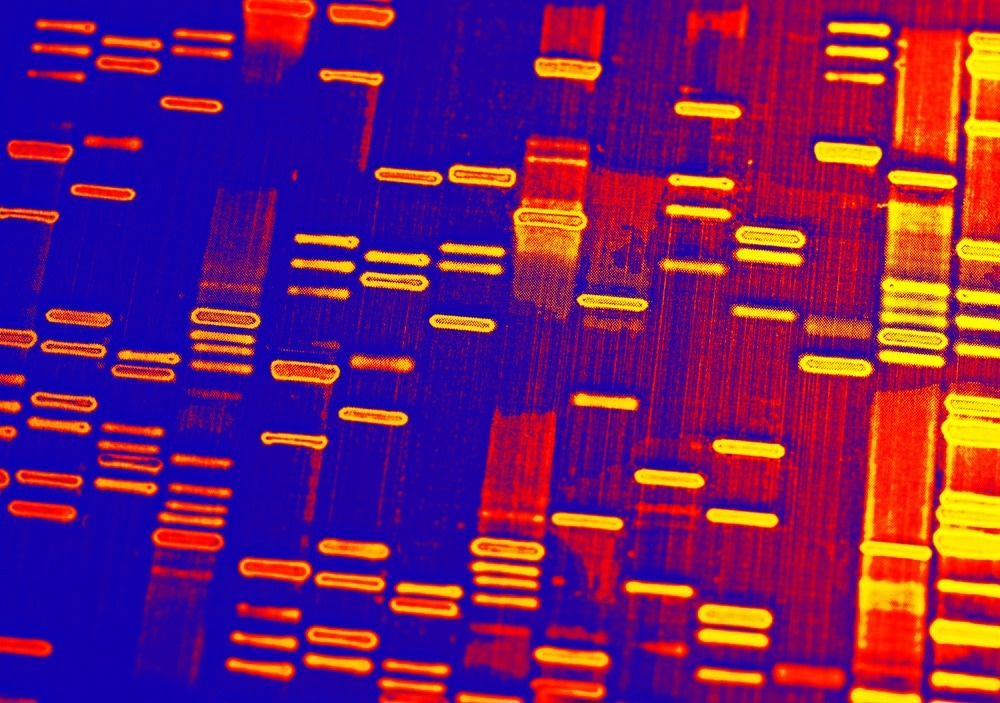
Image Credit: isak55/Shutterstock.com
Future Prospects
Although the application of CMOS systems in imaging cells is becoming more established, the technology also has a lot of potential in other fields. The use of CMOS systems in DNA sequencing equipment stands to be a promising area of development. The application of CMOS circuits in equipment such as Ion Torrent Systems has already been used in medical settings.
The development of smaller systems will increase the CMOS circuits application potential, although this is not the only improvement development companies are working towards. A decrease in power consumption and an increase in sensitivity and reliability are other important aims that will make the systems even more desirable.
The applications of this technology are numerous and have surpassed the methods currently in use in some respects. The increase in image quality, price, and size has made the CMOS systems incredibly desirable but still does not rival conventional microscopy. CMOS systems are a good alternative for field work, but more development is needed for it to be used as the default cell imaging technique.
References:
- Adiguzel, Y. & Kulah, H. (2012) CMOS Cell Sensors for Point-of-Care Diagnostics. Sensors. https://doi.org/10.3390/s120810042
- Christensson, P. (2017). CMOS Definition. [Online] TechTerms. Available at: https://techterms.com/definition/cmos (Accessed on 2 February 2022)
Last Updated: Feb 3, 2023

Written by
Grace Plahe
Grace is currently studying for her masters by research at the University of Salford, UK. Her research is looking into the advantages conferred by temperate bacteriophage to the Liverpool epidemic strain (LES) ofPseudomonas aeruginosain different conditions. This has applications in the control and treatment of cystic fibrosis infections. She is due to finish her project by the beginning of 2023 as she is studying part time while also working as a research technician.
Source: Read Full Article