Clinicians at the Smidt Heart Institute at Cedars-Sinai Medical Center in Los Angeles have developed a new algorithm that can effectively identify and distinguish between two cardiovascular conditions, hypertrophic cardiomyopathy and cardiac amyloidosis, that are often difficult to diagnose.
In a cohort study that included more than 23,000 patients, the algorithm, or deep-learning workflow, accurately identified subtle changes in left ventricular wall geometric measurements and the causes of hypertrophy.
The findings were published online in JAMA Cardiology.
“Increasingly, there is evidence that hypertrophic cardiomyopathy and cardiac amyloidosis are, although still rare, more common than we realized, but physicians often miss these conditions in the course of a routine diagnostic workflow,” senior author David Ouyang, MD, a cardiologist at Smidt Heart, told theheart.org | Medscape Cardiology.
“What we have done is automated the workflow to identify patients who likely have cardiac amyloidosis or hypertrophic cardiomyopathy. Our algorithm can zero in on disease patterns that can’t be seen by the naked eye, and then use these patterns to predict the right diagnosis,” Ouyang said.

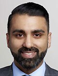
Dr David Ouyang
The cohort study, conducted from 2008-2020, included physician-curated cohorts from the Stanford Amyloid Center, Cedars-Sinai Medical Center (CSMC) Advanced Heart Disease Clinic for cardiac amyloidosis, the Stanford Center for Inherited Cardiovascular Disease, and the CSMC Hypertrophic Cardiomyopathy Clinic for hypertrophic cardiomyopathy.
The deep-learning algorithm was trained and tested on approximately 24,000 cardiac ultrasound videos from Cedars-Sinai and Stanford Healthcare’s echocardiography laboratories.
“When we look at heart ultrasounds, we are looking for features of the disease that can be easily overlooked. The two things that the algorithm looks for here are thick heart walls and unusual texture of the heart muscle, which can be detected on the ultrasound. The algorithm has been developed to identify those patients who should be seen by a clinician, so it efficiently flags those patients who are suspicious for having these conditions,” he said.
“We compared what the algorithm said were the patients most likely to have one of these conditions, and then compared the results with the patients’ actual records, and when we looked through, we found that the patients pointed out by the algorithm did in fact actually have one of the diseases. So, our algorithm identified high-risk patients with more accuracy than the well-trained eye of a clinical expert,” Ouyang said.
“I find the literature very exciting,” said Sumeet Mitter, MD, director of HFpEF and Cardiac Amyloidosis programs at Mount Sinai Hospital, New York City, who was asked to comment on this study.

Dr Sumeet Mitter
“It offers an ability to facilitate the recognition of these underdiagnosed ailments affecting the heart, including cardiac amyloidosis, in a streamlined manner,” said Mitter, who was not involved with the research.
“The strength of the study lies in the fact that the investigators used very large data sets from Stanford and Cedars-Sinai, two centers that are leading experts in the diagnosis of cardiac amyloidosis,” he told theheart.org | Medscape Cardiology.
The algorithm will have to be validated with echocardiograms from multiple institutions, perhaps even in a community setting, to see how it performs, Mitter suggested.
“The authors allude to that in their paper,” he noted. “We will need to validate this in a real-world setting, where data may not be acquired in as rigorous a fashion and in less-than-ideal settings. Eventually you want to be able to use this in the community as well, so it is not held only in tertiary medical centers.”
JAMA Cardiol. Published online February 28, 2022. Full text
For more from theheart.org | Medscape Cardiology, join us on Twitter and Facebook
Source: Read Full Article